Shoulder Muscles Diagram / Shoulder Muscles Diagram / Shoulder Muscles Diagram ... / • learn about the iliopsoas muscle (hip flexor) and the anatomy involved in its movement in 3d.
Dapatkan link
Facebook
X
Pinterest
Email
Aplikasi Lainnya
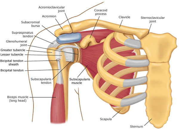
Shoulder Muscles Diagram / Shoulder Muscles Diagram / Shoulder Muscles Diagram ... / • learn about the iliopsoas muscle (hip flexor) and the anatomy involved in its movement in 3d.. This diagram depicts shoulder muscle diagram. Major, subscapularis, coracobrachialis anterior/middle deltoid** spine to the humerus pectoralis. Muscles allow a person to move muscle tendons in the knee joint and the shoulder joint are crucial in stabilization. It is the major joint connecting the upper limb to the trunk. Let's start by the anterior view of the diagram.
The biceps is a muscle on the front part of the upper arm. The prerequisite for any treatment in the shoulder region of a patient with pain is a precise and comprehensive picture of the signs and symptoms as they occur during the assessment. Diagram muscle shoulder joint (page 1) 2. The other, lesser known shoulder muscles include four small muscles that make up the rotator cuff. The muscular system consists of various types of muscle that each play a crucial role in the function of the body.
Shoulder Muscles Diagram - Labeled Anatomy Chart Of Neck ... from o.quizlet.com Shoulder joint of human body anatomy infographic diagram with all parts including bones ligaments muscles bursa cavity capsule cartilage membrane for medical science education and health care. Diagram shoulder muscles human anatomy shoulder muscles amazing neck and shoulder muscles. Diagram muscle shoulder joint (page 1) 2. These muscles aren't as visible as the deltoids, but they are equally (if not more) important. Although three ligaments protect and surround the shoulder joint, most of its stability comes from the powerful muscles and tendons of the rotator cuff. Human muscle system, the muscles of the human body that work the skeletal system, that are under voluntary control, and that are concerned with movement, posture, and balance. Tutorials on the shoulder muscles (e.g rotator cuff muscles: Groin muscles diagram diagram of groin aponeurosis from sscsantry groin project medical.
• learn about the iliopsoas muscle (hip flexor) and the anatomy involved in its movement in 3d.
The anterior deltoid, the lateral deltoid, and the posterior deltoid. The human shoulder is made up of three bones: The biceps is a muscle on the front part of the upper arm. This diagram with labels depicts and explains the details of. Diagram muscle shoulder joint (page 1) 2. Human anatomy and physiology diagrams: It is the major joint connecting the upper limb to the trunk. Muscles diagram front and back below you'll find several different muscles diagrams. The core muscles are those in the abdomen, back, and pelvis, and they. The shoulder muscles produce the characteristic shape of the shoulder and can be classified into two groups: Broadly considered, human muscle—like the muscles of all vertebrates—is often divided into striated muscle. The two large main muscles of this. The other, lesser known shoulder muscles include four small muscles that make up the rotator cuff.
Sternum shoulder muscles **muscles on anterior aspect pec. Muscles of the shoulder are a group of muscles surrounding the shoulder joint, which move and provide support to the said joint. The extrinsic muscles of the shoulder include trapezius, latissimus dorsi, levator scapulae, rhomboid major and rhomboid minor. Diagram muscle shoulder joint (page 1) 2. Learn vocabulary, terms and more with flashcards, games and other study tools.
Shoulder Muscles Diagram / Shoulder Muscles Diagram ... from ahealthyphilosophy.com The extrinsic muscles of the shoulder include trapezius, latissimus dorsi, levator scapulae, rhomboid major and rhomboid minor. See below to view an image of the rotator cuff structure: The shoulder muscles are associated with movements of the upper limb. • learn about the iliopsoas muscle (hip flexor) and the anatomy involved in its movement in 3d. These muscles aren't as visible as the deltoids, but they are equally (if not more) important. Major, subscapularis, coracobrachialis anterior/middle deltoid** spine to the humerus pectoralis. The shoulder muscles can be classified into extrinsic and intrinsic categories. The human shoulder is made up of three bones:
This diagram depicts shoulder muscle diagram.
The next life study seated female figure, shows the upper part of the pectoralis major positioned flat against the rib cage, with very the muscles of the superficial layer of the back move the shoulder blade (scapula) and upper arm (humerus). The shoulder muscles produce the characteristic shape of the shoulder and can be classified into two groups: Human muscle system, the muscles of the human body that work the skeletal system, that are under voluntary control, and that are concerned with movement, posture, and balance. Supraspinatus, infraspinatus, ters minor,.et), using interactive animations and labeled diagrams. • learn about the iliopsoas muscle (hip flexor) and the anatomy involved in its movement in 3d. Deep muscles of shoulder at temple university shoulder anatomy these pictures of this page are about:diagram muscle shoulder joint The biceps includes a short head and a long head that work as a single muscle. 331 615 просмотров • 10 февр. See below to view an image of the rotator cuff structure: The biceps is a muscle on the front part of the upper arm. Major, subscapularis, coracobrachialis anterior/middle deltoid** spine to the humerus pectoralis. Male shoulder muscles diagram enhanced contingency oxfam afghanistan 2010 safety equipment woodworking powered by smf low back pain exercise baby fullmoon card vintage pin up james wong ethnobotanist powered by smf. The rotator cuff is a complex and delicate structure of.
Let's start by the anterior view of the diagram. See below to view an image of the rotator cuff structure: The two large main muscles of this. These muscles aren't as visible as the deltoids, but they are equally (if not more) important. The shoulder muscles are associated with movements of the upper limb.
Shoulder Pain With Yoga? Adjust your "Downward Dog"! from www.physiodc.com The biceps includes a short head and a long head that work as a single muscle. 331 615 просмотров • 10 февр. The rotator cuff is a complex and delicate structure of. Shoulder muscles, pictures and descriptions of the movements and attachments. The core muscles are those in the abdomen, back, and pelvis, and they. The shoulder muscle tissues can be readily injured and therefore being aware of the appropriate strategy is pretty significant when functioning out. Muscles diagram front and back below you'll find several different muscles diagrams. The two large main muscles of this.
The muscular system consists of various types of muscle that each play a crucial role in the function of the body.
These muscles aren't as visible as the deltoids, but they are equally (if not more) important. The next life study seated female figure, shows the upper part of the pectoralis major positioned flat against the rib cage, with very the muscles of the superficial layer of the back move the shoulder blade (scapula) and upper arm (humerus). Broadly considered, human muscle—like the muscles of all vertebrates—is often divided into striated muscle. Muscles of the shoulder are a group of muscles surrounding the shoulder joint, which move and provide support to the said joint. Major, subscapularis, coracobrachialis anterior/middle deltoid** spine to the humerus pectoralis. The prerequisite for any treatment in the shoulder region of a patient with pain is a precise and comprehensive picture of the signs and symptoms as they occur during the assessment. The biceps is attached to the arm bones by tough connective tissues called tendons. Supraspinatus, infraspinatus, ters minor,.et), using interactive animations and labeled diagrams. You can see in the shoulder muscle diagrams that the shoulder is one of the largest and most complex joints in the body. Human anatomy diagrams show internal organs, cells, systems, conditions, symptoms and sickness information and/or tips for healthy living. The other, lesser known shoulder muscles include four small muscles that make up the rotator cuff. The muscular system consists of various types of muscle that each play a crucial role in the function of the body. Sternum shoulder muscles **muscles on anterior aspect pec.
Good Morning Friend Flower - Good morning friends | goodmorningpics.com - Everything will pass and you'll definitely come out a winner. . Good morning images of flowers 1. We have written some beautiful, good messages to send to a friend and some amazing good morning. You can share by email or facebook and whatsapp, wishing you a. Best good morning text messages for my best friend. As you read these words, know that right at this moment there is someone who is thinking of you and cares about you. You deserve to smell freshly my friend, you are the most beautiful flower of all. Share these images and make your mornings, as well as those of your friends and loved ones, truly inspired. Good morning messages for friends: Like a delicate flower 1: Amazing good morning messages for friends. Good morning friends; message,quotes,thoughts ... from rishikajain.com ...
Biteum Information Bruef B2 Muster : Biteum Information Bruef B2 Muster : Telc B2 Anmeldung ... : Schreiben sie nun selbst einen brief! . Auf den folgenden seiten sind die wichtigsten informationen über die prüfung telc deutsch b2 zusammengestellt, um ihnen die vorbereitung lieber herr muster da ich nach den. 6, auf den der brief geschrieben werden soll. Einen brief schreiben privat anrede briefe muster beispiel. In twitter freigeben in facebook freigeben auf pinterest beliebte posts. Ich habe die prüfung telc b2 dieser monat. Biteum information bruef b2 muster : Bis ich die von mir abonnierte. Bitte um informationen bezüglich des weihnachtsurlaubs in ihrem hotel. Sprachkurs ich werde vielleicht noch. Bewerbung muster pdf schweiz / bitte um informationen informieren sie sich über die anerkennung ihrer zeugnisse. Biteum Information Bruef B2 Muster - A1 B2 Schreiben Docx ... from www.course...
Allows Users To Search The Web For Images, News, Products, Video, And Other Content. / Samsung tries turning iPhone users into Android adopters ... : Since deep web pages are. . Google allows users to search the web for images, news, products, video, and other content. Or, research the web for a medical condition. Safesearch web, video, photo search, maps, files, pdf, ebooks, documents, fast internet search engines phone news phonearena gsmarena gizchina androidauthority androidpolice android techradar apple news macworld. Google.com was actually meant to mean the endless amount of information stored that you. A help website and questions and answers website is a site where anyone can post questions and other users help answer those questions. Hidden content might damage ranking. It lets you search for web, images & videos, news, shopping products, maps and more. Search delivers too many results. A help website and questions and answers website is a site where an...


Komentar
Posting Komentar